
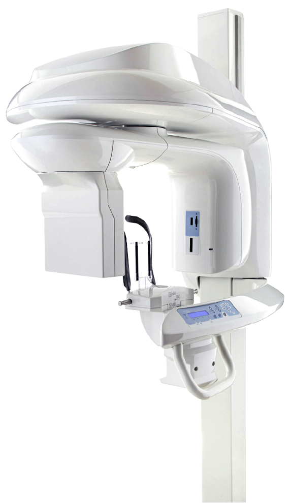
3D Cone Beam CT Imaging
Our oral and maxillofacial surgeons use 3D cone beam computed tomography (CBCT) when additional imaging is helpful for diagnosis or surgical planning. CBCT provides detailed three-dimensional views of the teeth, jaws, facial bones, nerves, sinuses, and surrounding structures that may not be fully visible on traditional two-dimensional dental X-rays.
What Is a Cone Beam CT Scan?
A cone beam CT scan uses a rotating X-ray source to capture multiple images from different angles. Specialized software then combines these images to create a three-dimensional representation of the area being examined.
Unlike a conventional medical CT scan, dental CBCT is designed specifically for imaging the teeth, jaws, and facial structures. The area being scanned can be selected according to the information needed for an individual patient’s evaluation or treatment.
How We Use 3D Imaging in Oral Surgery
CBCT imaging can provide important information for many oral and maxillofacial surgery procedures. Depending on the individual situation, our surgeons may use 3D imaging to assist with:
- Dental implant planning — evaluating available bone and the location of nearby anatomical structures before implant placement.
- Wisdom teeth and impacted teeth — evaluating the position of impacted teeth and their relationship to surrounding teeth, bone, nerves, or other structures.
- Bone grafting — assessing the amount and shape of available bone when planning grafting or reconstructive procedures.
- Sinus augmentation — evaluating the maxillary sinus and surrounding bone when planning certain upper-jaw dental implant procedures.
- Oral pathology — evaluating selected cysts, lesions, or other abnormalities involving the jaws and surrounding structures.
- Facial and jaw conditions — providing three-dimensional information that may assist in evaluating facial injuries, jaw abnormalities, or other surgical conditions.
3D Imaging for Dental Implant Planning
Three-dimensional imaging can be particularly useful when planning dental implant surgery. A CBCT scan allows the surgeon to evaluate the height and width of the available bone as well as the location of structures such as nerves and the maxillary sinuses.
This information can help determine an appropriate implant position and whether procedures such as bone grafting or sinus augmentation may need to be considered as part of treatment planning.
Do All Oral Surgery Patients Need a CBCT Scan?
No. Three-dimensional imaging is not necessary for every patient or every procedure. Your surgeon will determine what type of imaging is appropriate based on your examination, existing dental X-rays, medical and dental history, and the treatment being considered.
If adequate recent imaging has already been obtained by your dentist or another healthcare provider, please let our office know. When appropriate, those images may be reviewed as part of your evaluation.
Coordinating With Your Dentist or Other Providers
Digital imaging also allows our surgeons to review anatomical findings carefully and, when appropriate, coordinate treatment with your general dentist, orthodontist, prosthodontist, physician, or other healthcare provider.
Our goal is to use imaging when it provides meaningful information for diagnosis, treatment planning, or communication about your care.